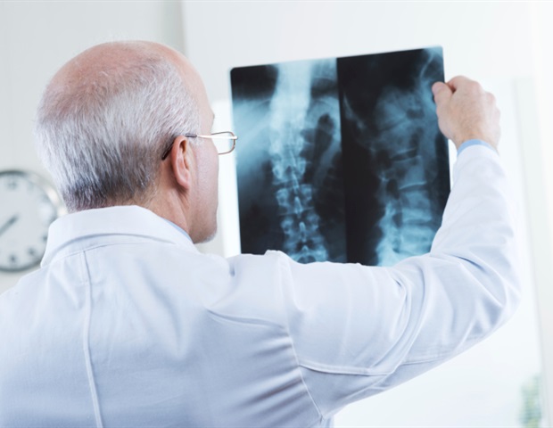
The prognosis for recovery from a spinal cord injury (SCI) is of great importance for those directly affected and those around them. So far, however, it has remained imprecise. Researchers from three international rehabilitation centers in Zurich, Murnau and Denver have now succeeded in demonstrating the value of neuroimaging measurements for predicting sensory and motor recovery in people with quadriplegia.
Neuroimaging measurements derived from clinical magnetic resonance imaging (MRI) record the extent of the uninjured nerve tissue next to the spinal cord lesion, known as "spinal tissue bridges". The results of the longitudinal study "Prognostic value of tissue bridges in cervical spinal cord injury" have the potential to change clinical practice. They have just been published in The Lancet Neurology, the world's leading journal of clinical neurology.

The team led by lead author Dr. Dario Pfyffer and senior author Prof. Dr.
med. Patrick Freund from Balgrist University Hospital and the University of Zurich, which includes SCI experts from around the world, has successfully developed models that incorporate tissue bridges in the spinal cord in a large, multicenter cohort of patients with cervical SCI for improved prognosis of clinical outcomes. These tissue bridges were measured on MRI images (taken early after the onset of the spinal cord injury).
This has resulted in significant added value for the previous prognosis models, which are based on recording the clinical condit.