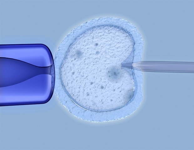
Innovative research, presented today at the ESHRE 40 th Annual Meeting in Amsterdam, has introduced a novel 3D imaging model designed to identify features of blastocysts – the early stage of development for an implanted embryo – associated with successful pregnancies. This new approach could transform current blastocyst selection methods, and open avenues for increased pregnancy rates. The shape and structure of blastocysts can predict the success of a pregnancy, aiding blastocyst selection for in vitro fertilization (IVF).
However, selecting the right embryo or blastocyst remains a significant challenge within IVF. Traditionally, the quality of blastocysts is assessed using 2D methods that lack depth and comprehensive indicators. Although some 3D methods exist, they aren't practical or safe for clinical use.

This study bridges that gap by introducing a clinically applicable 3D evaluation method and reveals previously unrecognized spatial features of blastocysts indicative of outcomes." Dr. Bo Huang, lead author of the study The study included women under 40 years old with a uterine lining (endometrial thickness) of 7-16mm and no more than one previous embryo transfer failure.
Using a device called EmbryoScope+, researchers took detailed images of 2,141 frozen-thaw single blastocysts. Advanced technology was used to create 3D models of these blastocysts, capturing detailed information about their outer layer (trophectoderm) and inner cell mass. These models were further a.