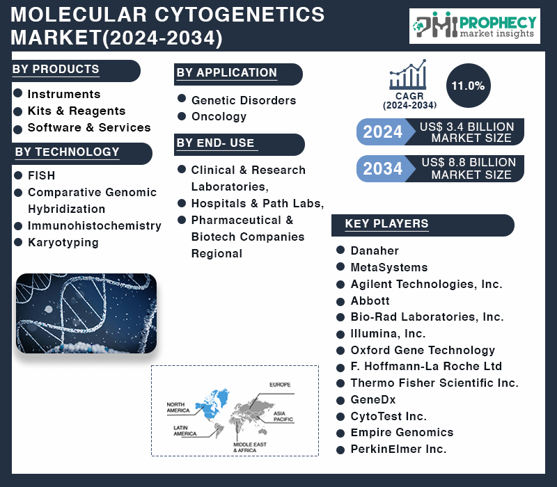
Covina, June 26, 2024 (GLOBE NEWSWIRE) -- Molecular cytogenetics is a field that combines molecular biology and cytogenetics to analyze chromosome structure to distinguish normal and cancer-causing cells. It began in 1956 with the discovery of normal human cells having 46 chromosomes. The field is now used to diagnose and treat various malignancies, including hematological malignancies and brain tumors.
Techniques like fluorescence in situ hybridization (FISH) are used to visualize specific DNA regions and assess copy number changes in the entire genome. Fluorescence In Situ Hybridization (FIH) is a technique used to map out single copies or repetitive DNA sequences through localization labeling of specific nucleic acids. It uses different DNA probes labeled with fluorescent tags to bind to specific regions of the genome.

FISH can be performed on interphase cells and paraffin block tissues and can be performed directly or indirectly. Comparative genomic hybridization (CGH) is a method used to compare variations in copy number between a biological sample and a reference. It uses two genomes, a sample, and a control, labeled fluorescently to distinguish them.
CGH can point to gains or losses of chromosomal regions and can scan an entire genome quickly for chromosome imbalances. Array comparative genomic hybridization (aCGH) allows CGH to be performed without cell culture and isolation, using glass slides containing small DNA fragments. This process allows for thousands of signa.