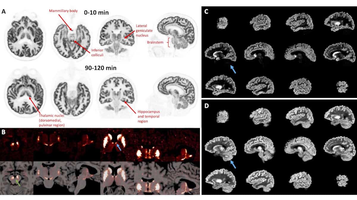
A series of ultrahigh-resolution brain PET images has been selected as the SNMMI Image of the Year. At each of its annual meetings, the Society of Nuclear Medicine and Molecular Imaging chooses an image that represents the most promising advances in the field, with this year’s winner picked from more than 1500 submitted abstracts. The winning images, recorded by the ultrahigh-performance NeuroEXPLORER human brain PET scanner, highlight targeted tracer uptake in specific brain nuclei (clusters of neurons), providing detailed information on neuronal and functional activity.
The technology could dramatically expand the scope of brain PET studies, with potential to advance the treatment of brain diseases. NeuroEXPLORER was built by a collaboration of researchers from Yale University , the University of California, Davis and United Imaging Healthcare . They designed the scanner to provide ultrahigh sensitivity and ultrahigh spatial resolution, as well as to perform continuous correction for head motion.

“If we are going to get high resolution, we have to deal with patient motion,” Richard Carson from Yale University explained at the recent SNMMI Annual Meeting in Toronto. “But the challenge has always been sensitivity, even with a high-resolution system. In a late frame for a carbon-11 study, you’re really scanning on fumes, it’s hard to generate the quality of images you’d like to really use the resolution that’s possible.
” To achieve the highest possible resolu.


