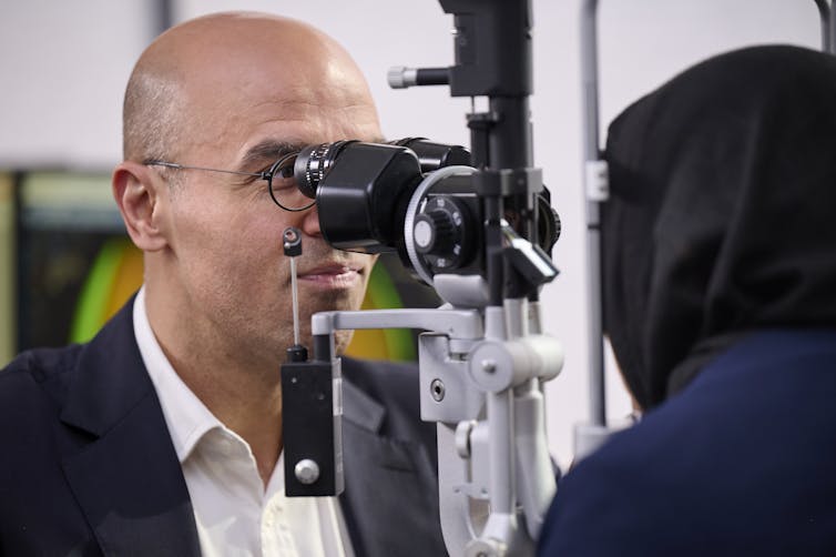
My first encounter came as a medical student. Under high magnification, I examined a colleague’s iris, the coloured part of their eye encircling the pupil. I watched as the muscle fibres moved rhythmically, undulating between dilation and constriction.
It looked like an underwater plant, swaying in a current. Mesmerising. But in a busy university curriculum the experience quickly faded, to be replaced by the next clinical rotation.

I forgot ophthalmology; “maybe orthopaedic surgery or emergency medicine are for me”, I thought. But eyes returned, this time while I was a junior doctor in residency. Assisting in surgery, I observed a patient’s retina through an operating microscope.
Here was a cinematic view of the orb, as if viewed from a spacecraft over a Martian landscape. The internal surface glowed blood orange (the colour once ascribed to its rich blood supply, now attributed to a layer of underlying pigmented cells). Within this landscape ran red rivulets, a network of branching blood vessels.
The Greek anatomist Herophilus thought this pattern resembled a casting net, leading to “retiform” (meaning reticular or netlike), which became “retina” in contemporary language (the light-sensitive film at the back of the eye). I was struck by the intricacy of this secret globe, this gallery of miniature art. The term “beauty is in the eye of the beholder” took on new connotations, and I turned to pursuing ophthalmology.
Aside from the organ’s intrinsic appeal.